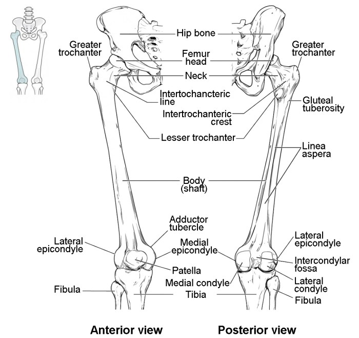
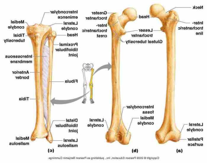
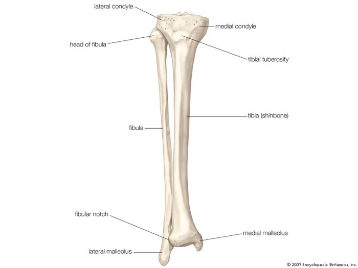
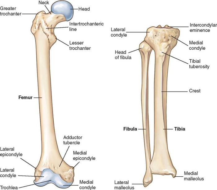
Label the bones and bone markings of the lower leg, embarking on an anatomical journey that unravels the intricate structure and function of this essential part of the human body. From the tibia’s pivotal role in weight-bearing to the fibula’s contribution to ankle stability, this exploration delves into the fascinating world of skeletal anatomy.
The narrative unfolds, meticulously detailing the medial and lateral condyles, tibial tuberosity, and anterior crest, highlighting their significance in movement and stability. The fibula’s fibular head, lateral malleolus, and soleal line emerge as crucial landmarks, underscoring their clinical relevance.
Lower Leg Bones: Label The Bones And Bone Markings Of The Lower Leg

The lower leg, also known as the crus, consists of two long bones: the tibia and the fibula. These bones provide structural support, facilitate movement, and protect vital structures within the leg.
Tibia
The tibia, or shinbone, is the larger and more medial of the two bones. It extends from the knee joint to the ankle joint and bears the majority of the body’s weight.
The tibia has a triangular cross-section, with a broad medial surface and a narrower lateral surface. The proximal end of the tibia features two condyles: the medial and lateral condyles. These condyles articulate with the condyles of the femur, forming the knee joint.
The anterior surface of the tibia is characterized by the tibial tuberosity, a roughened area where the patellar ligament attaches. The tibial tuberosity provides leverage for knee extension.
The lateral surface of the tibia features the anterior crest, a ridge that extends from the proximal to the distal end of the bone. The anterior crest serves as an attachment site for muscles that control ankle movement.
Fibula, Label the bones and bone markings of the lower leg
The fibula is a slender bone located lateral to the tibia. It extends from the knee joint to the ankle joint but does not bear any weight.
The proximal end of the fibula is expanded to form the fibular head, which articulates with the lateral condyle of the tibia. The distal end of the fibula forms the lateral malleolus, a bony prominence on the outer side of the ankle joint.
The fibula serves several important functions. It provides stability to the lower leg, prevents excessive inward movement of the ankle, and acts as an attachment site for muscles that control foot and ankle movement.
Questions Often Asked
What is the primary function of the tibia?
The tibia is the larger and weight-bearing bone of the lower leg, providing structural support and facilitating movement.
What is the significance of the soleal line on the fibula?
The soleal line serves as an attachment point for the soleus muscle, contributing to ankle plantarflexion and stability.


