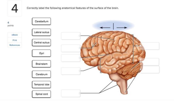
Correctly label the following anatomical features of the eye. – Embark on a journey to accurately identify the intricate anatomical components of the eye. From the cornea’s transparent layers to the lens’s focusing capabilities, this exploration unveils the remarkable intricacies of our visual system.
Prepare to delve into the depths of ocular anatomy, unraveling the functions and structures that orchestrate our perception of the world around us.
Corneal Structure

The cornea is the transparent, dome-shaped outermost layer of the eye that covers the pupil, iris, and anterior chamber. It is composed of five distinct layers:
- Epithelium: The outermost layer, consisting of non-keratinized stratified squamous epithelium, protects the cornea from external factors.
- Bowman’s Layer: A thin, acellular layer that provides additional strength and support to the cornea.
- Stroma: The thickest layer, consisting of collagen fibers arranged in a regular lattice pattern, providing transparency and structural integrity.
- Descemet’s Membrane: A thin, elastic layer that acts as a barrier between the stroma and the endothelium.
- Endothelium: A single layer of flat, hexagonal cells that pumps excess fluid out of the cornea, maintaining its transparency.
The cornea’s transparency is essential for clear vision. Its curvature helps refract light, directing it towards the lens and retina.
Iris Function
The iris is a thin, muscular diaphragm that gives the eye its color and controls the size of the pupil. It is composed of two muscles:
- Sphincter pupillae: A circular muscle that constricts the pupil.
- Dilator pupillae: A radial muscle that dilates the pupil.
The iris regulates the amount of light entering the eye by adjusting the size of the pupil. In bright light, the sphincter pupillae contracts, narrowing the pupil to reduce light intensity. Conversely, in dim light, the dilator pupillae relaxes, widening the pupil to allow more light to enter.The
variation in iris color is determined by the amount and distribution of melanin, a pigment produced by melanocytes in the iris stroma.
Lens Anatomy
The lens is a transparent, biconvex structure located behind the iris and pupil. It is composed of three main parts:
- Lens capsule: A thin, elastic membrane that surrounds the lens.
- Lens cortex: The bulk of the lens, consisting of lens fibers arranged in concentric layers.
- Lens nucleus: The central, dense core of the lens.
The lens’s primary function is to focus light onto the retina. It changes its shape through the action of the ciliary body, a ring of muscles that surrounds the lens. By adjusting its curvature, the lens fine-tunes the focal length, ensuring clear vision at various distances.
Retina Structure
The retina is a thin, light-sensitive layer that lines the back of the eye. It is composed of several layers, each with specific functions:
- Retinal pigment epithelium: A layer of pigmented cells that absorbs excess light, preventing its reflection within the eye.
- Photoreceptor layer: Contains two types of photoreceptor cells, rods and cones, which convert light into electrical signals.
- Outer nuclear layer: Contains the cell bodies of photoreceptors.
- Outer plexiform layer: Where photoreceptor synapses connect with bipolar cells.
- Inner nuclear layer: Contains the cell bodies of bipolar and horizontal cells.
- Inner plexiform layer: Where bipolar and horizontal cells synapse with ganglion cells.
- Ganglion cell layer: Contains the cell bodies of retinal ganglion cells, which transmit visual information to the brain via the optic nerve.
- Nerve fiber layer: Contains the axons of retinal ganglion cells, which form the optic nerve.
The macula and fovea are specialized regions of the retina responsible for central vision and high-acuity color perception.
Optic Nerve Pathway, Correctly label the following anatomical features of the eye.
The optic nerve is a bundle of nerve fibers that transmits visual information from the retina to the brain. It follows a complex pathway:
- Retina: The optic nerve fibers originate from retinal ganglion cells.
- Optic chiasm: The optic nerves from both eyes partially cross at the optic chiasm, forming the optic tracts.
- Optic tracts: The optic tracts continue towards the brain.
- Lateral geniculate nucleus: The optic tracts terminate in the lateral geniculate nucleus of the thalamus, where visual information is processed.
- Optic radiations: From the lateral geniculate nucleus, the visual information is relayed to the visual cortex in the occipital lobe of the brain.
The optic nerve pathway is crucial for transmitting visual signals to the brain for interpretation and conscious perception.
Query Resolution: Correctly Label The Following Anatomical Features Of The Eye.
What is the function of the cornea?
The cornea is the transparent outermost layer of the eye that serves as a protective barrier and plays a crucial role in refracting light, focusing it onto the retina.
How does the iris control pupil size?
The iris, the colored part of the eye, contains muscles that contract and relax, adjusting the size of the pupil to regulate the amount of light entering the eye.
What is the role of the lens in the eye?
The lens is a flexible structure located behind the iris that changes shape to focus light onto the retina, ensuring clear vision at various distances.

